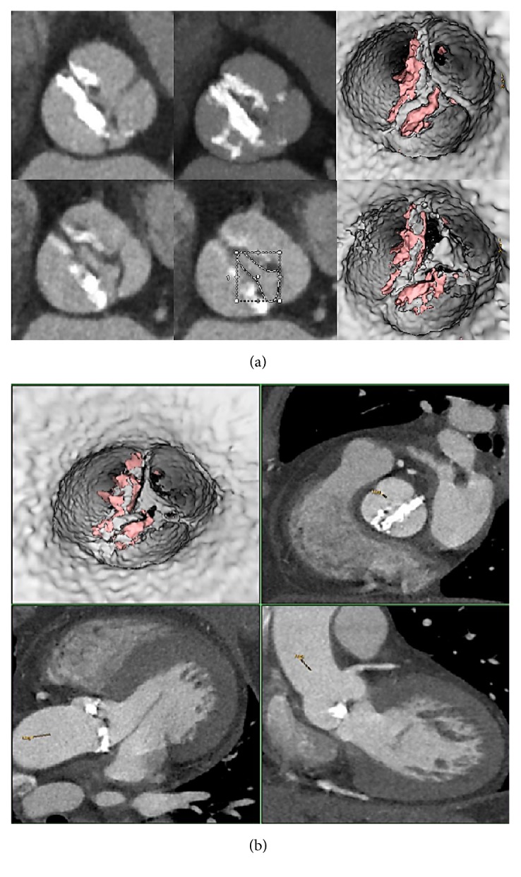
Figure 1.

320 CT scan shows refractory calcifications on aortic leaflets of a prohibitive high-risk patient who undergo TAVR. The procedure performed was as follows: one beat full cardiac cycle (0-100%) acquisition DLP = 459 mGy/cm for functional aortic valve assessment, morphological aortic valve study, and anatomical AVA determination.
