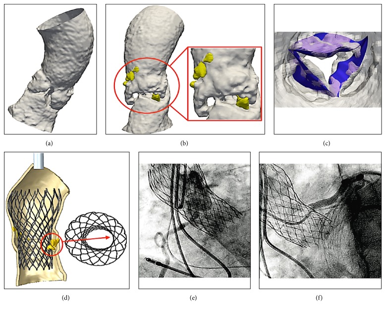
Figure 8.
FEA of aortic root and leaflets from CT images after TAVR (a, b, c, d, and e). Calcific blocks and segmentation of blood flow show jagged surface of Valsalva sinus and leaflets (a, b, c). Bulky calcifications determine a noncomplete stent bottom expansion (d). The migration of the device causes the thrombosis of the coronary ostia (e, f).

