Figure 3.
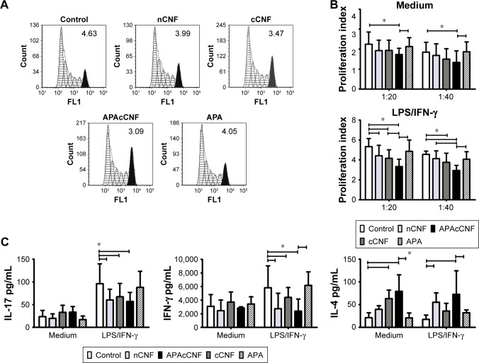
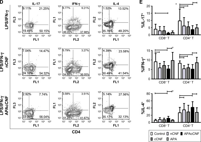
Effects of CNF samples on Th-polarization capacity of DC.
Notes: (A) A representative analysis of CFSE-labeled CD3+ T-cell proliferation in coculture with LPS/IFN-γ stimulated DC at 1:20 (DC:T cell) ratio (the number indicate proliferation index). (B) The summarized results on proliferation from four independent experiments are shown as mean proliferation index ± SD. (C) The levels of cytokines in DC/CD3+ T-cell cocultures measured by specific ELISA are shown as mean pg/mL ± SD from four independent experiments. (D) A representative analysis of intracellular cytokines’ expression in CD3+ T cells from DC/T cocultures. The data from three experiments with T cells which showed a marked expression of cytokines are shown. This was done since highly variable production of these cytokines by different T-cell donors was shown previously.45 (E) The summarized data from five independent experiments carried out with LPS/IFN-γ-stimulated DC are shown as mean % of positive cells within CD4+ or CD4− (CD8+) T cells ± SD. *P<0.05 as indicated (RM ANOVA).
Abbreviations: APA, 3-aminopropylphosphoric acid; APAc, APA-functionalized; c, carboxylated; CFSE, carboxyfluorescein succinimidyl ester; CNF, cellulose nanofibrils; DC, dendritic cells; IFN, interferon; LPS, lipopolysaccharide; n, native; RM, repeated measures; Th, T helper cell.


