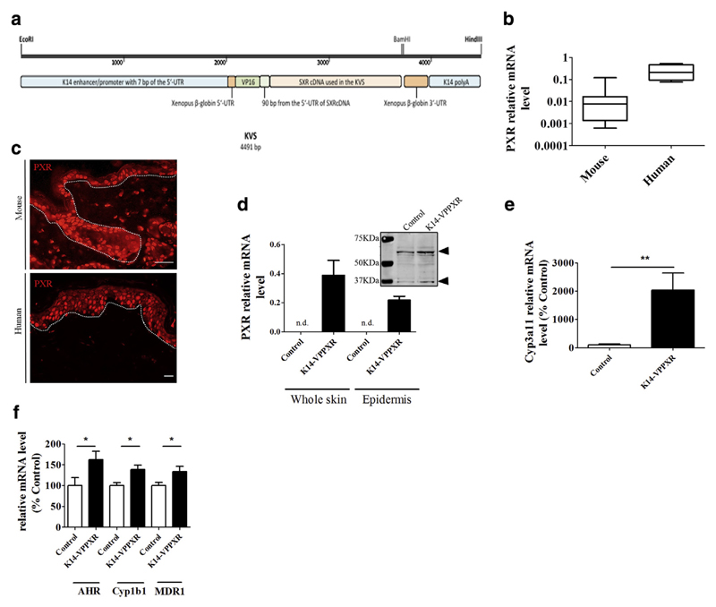
Figure 1. PXR locates to basal keratinocytes in mouse skin, and constitutive activation of PXR in the epidermis leads to an overall upregulation of xenobiotic metabolism.
(a) The KVS transgene (drawn to scale) depicting the human SXR cDNA attached to the VP16 domain sequence as subcloned in the K14 cassette. The VP16 domain is from herpes simplex virus, and the K14 sequences are from the human K14 gene. The EcoRI-HindIII linear fragment was used to generate transgenic mice (see also Supplementary Materials and Methods and Supplementary Figure S1). (b) Quantitative PCR showing relative expression of PXR mRNA levels relative to housekeeping genes in mouse (n = 10) and human (n = 4) skin. (c) Immunohistochemistry showing PXR staining in mouse (upper panel) and human (lower panel) skin. Bar = 50 μm. Dotted lines separate the epidermis from the dermis. (d) Quantitative PCR showing relative expression of human PXR mRNA levels in the skin or the epidermis of control (n = 5) and K14-VPPXR (n = 12) mice. Western blot analysis shows mouse PXR in control mouse skin and mouse + transgenic PXR expression in the K14-VPPXR mouse skin (pools of three skins). Arrows indicate the PXR. Quantitative PCR shows relative expression of (e) Cyp3A11 (n = 5) and (f) Ahr, Cyp1B1, and Mdr1/Abcb1 (n = 10–19) in the K14-VPPXR and control mouse skin. Data were analyzed with a Student’s t-test. *P <0.05, **P < 0.01. K14, keratin 14; n.d., not detectable; PXR, pregnane X receptor.

