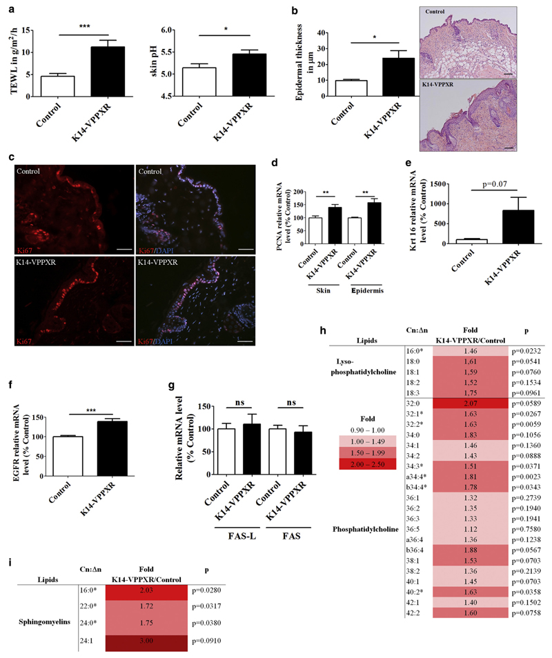
Figure 2. Constitutive activation of PXR in the epidermis leads to skin barrier defects and alters local homeostasis.
(a) TEWL (n = 29) and skin surface pH (n = 9–13) in adult K14-VPPXR and control mice. (b) Epidermal thickness and representative pictures of hematoxylin and eosin staining from the K14-VPPXR and control mouse back skin. Bar = 100 μm. (c) Representative images of Ki67 staining from K14-VPPXR and control mouse skin. Nuclei are counterstained with DAPI. Bar = 50 μM. Quantitative PCR showing relative expression levels of (d) Pcna, (e) Krt16, (f) Egfr, and (g) Fas and Fas-L in K14-VPPXR and control mouse epidermis or whole skin (n = 5–19). Fold increase and P values for epidermal (h) lysophosphatidylcholine and (i) sphingomyelin lipid species. Means of lipid amounts in K14-VPPXR mouse epidermis were divided by those in controls, n = 10–11. Data were analyzed with a Student's t-test: *P < 0.05, **P < 0.01, ***P < 0.001. K14, keratin 14; ns, nonsignificant; PXR, pregnane X receptor; TEWL, transepidermal water loss.

