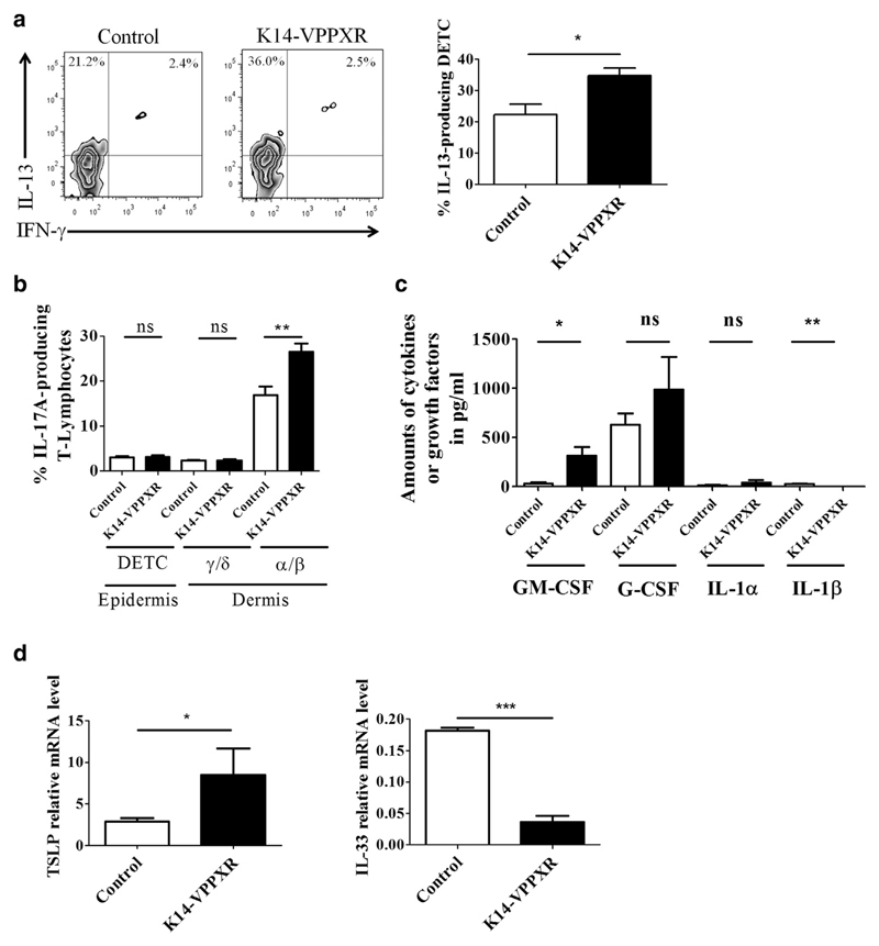
Figure 4. Sources of cytokines and growth factors in the skin of K14-VPPXR transgenic mice.
Epidermal and dermal cell suspensions were prepared as described in the Materials and Methods section and stained for flow cytometric analyses. All analyses used a pregate on CD45+ viable cells. (a) Representative FACS and percentages of IL-13-producing DETCs (CD4+TCRγ/δ+) in K14-VPPXR and control mouse epidermis, n = 5. (b) Percentages of IL-17A-producing skin-derived T lymphocytes (CD4+TCRγ/δ+ or CD4+TCRα/β+) in K14-VPPXR and control mouse skin, n = 5. (c) Production of cytokines by second-passage K14-VPPXR and control mouse keratinocytes analyzed by multi-analytical ELISA (n = 3). (d) Quantitative PCR showing relative expression of TSLP and IL33 in second-passage K14-VPPXR and control mouse keratinocytes. Three independent experiments were performed. Data were analyzed with a Student’s t-test. *P < 0.05, **P < 0.01, ***P < 0.0001. DETC, dendritic epidermal TCRγ/δ+ T cells; K14, keratin 14; ns, nonsignificant; PXR, pregnane X receptor.

