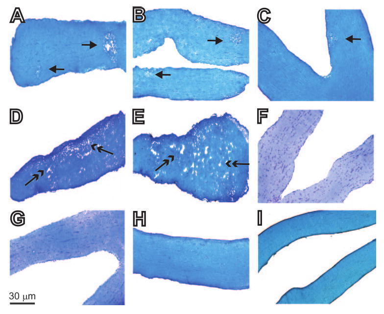
Figure 1.

Histologic analyses of optic nerves obtained from female BALB/c mice 14 days after ocular infection with HSV-IL-2 or control viruses. Female BALB/c mice were infected ocularly with HSV-IL-2, HSV-IL-4, HSV-IFN-γ, KOS, or mock-infected, as described in the footnote to Table 1. The optic nerves were obtained 14 days later, fixed, sectioned, and stained with LFB. Representative photomicrographs are shown. In the photomicrograph of the section of the optic nerve from the HSV-IL-2–infected mouse, the single-headed arrow indicates focal demyelination, and the two-headed arrow indicates diffuse demyelination. (A–C) In HSV-IL-2–infected optic nerve areas of focal demyelination were apparent as plaque areas with loss of LFB staining (single-headed arrow). (D, E) In HSV-IL-2–infected optic nerve, areas of diffuse demyelination were apparent as plaque areas with loss of LFB staining (two-headed arrow). In contrast to (A–E), no demyelination was detected in mice infected with HSV-1 strain KOS (F), HSV-IL-4 (G), HSV-IFN-γ (H), or mock-infected (I) as shown by dense LFB blue staining in myelinated regions, and no detectable focal or diffuse positive material.
