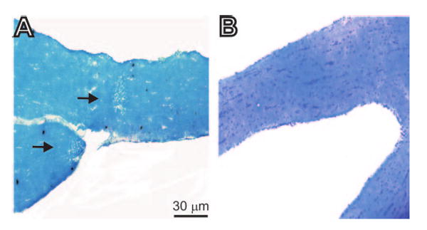
Figure 2.

Histologic analyses of optic nerves obtained from female BALB/c mice 10 days after ocular infection with HSV-IL-2 or control HSV-IL-4. Female BALB/c mice were infected ocularly with HSV-IL-2 or -IL-4, as described in the footnote to Table 1. The optic nerves were obtained 10 days later, fixed, sectioned, and stained with LFB. Representative photomicrographs are shown. (A) In HSV-IL-2–infected optic nerve, areas of focal demyelination were apparent as plaque areas (arrows). (B) No demyelination was detected in mice infected with HSV-IL-4.
