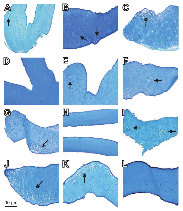
Figure 4.

Effect of ocular HSV-IL-2 infection on optic nerve demyelination in different strains of mice. Female C57BL/6, 129SVE, or SJL/6 mice were infected ocularly with HSV-IL-2 (2 × 10⁵ PFU/eye). Control mice were infected similarly with HSV-IL-4. The optic nerves were dissected 14 days later, fixed, sectioned, and stained with LFB. Representative micrographs are shown. In the photomicrograph of the section of the optic nerve from the HSV-IL-2–infected mouse, the single-headed arrow indicates focal demyelination, whereas the two-headed arrow indicates diffuse demyelination. (A–C) C57BL/6 mice infected with HSV-IL-2; (D) C57BL/6 mice infected with HSV-IL-4; (E–G) 129SVE mice infected with HSV-IL-2; (H) 129SVE mice infected with HSV-IL-4; (I–K) SJL/6 mice infected with HSV-IL-2; and (L) SJL/6 mice infected with HSV-IL-4.
