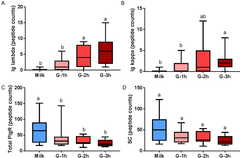
Fig. 5.

Peptide counts of immunoglobulin (Ig) fragments in human milk (blue boxplot) and preterm infant gastric samples at 1 (G-1h), 2 (G-2h) and 3 h (G-3h) after the beginning of feeding. Peptide counts of (A) Ig lambda (from IgA, SIgA, IgM, SIgM or IgG), (B) Ig kappa (from IgA, SIgA, IgM, SIgM or IgG), (C) total polymeric immunoglobulin receptor (PIgR) (f19–764) and (D) secretory component (SC, f19–603 of PIgR). Paired milk and gastric samples were collected in preterm-delivering mother-infant pairs (23–32 wk of gestational age (GA) and 7–98 days of postnatal age). Letters a, b and c show statistically significant differences between groups (p < 0.05) using one-way ANOVA with repeated measures followed by Tukey’s multiple comparison tests. Values are min, median and max, n = 11.
