
Figure 1: Inducible and conditional deletion of Ezh2 at E8.5 in both the CNCC-derived and PM- derived CM.
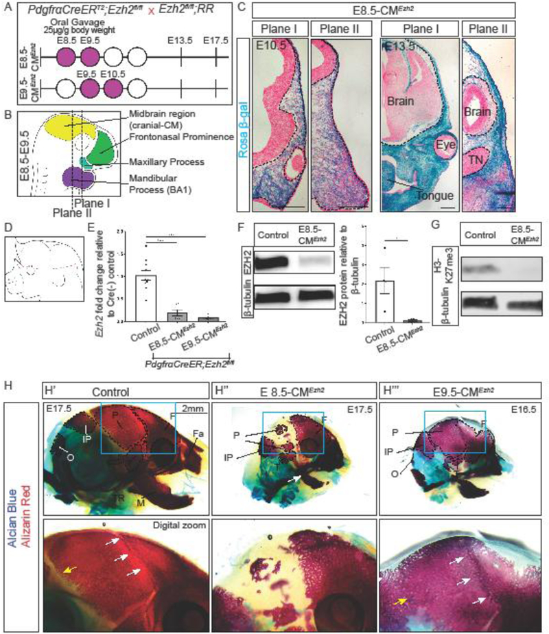
(A) Mating strategy and gavage regimen for conditional Ezh2 deletion for E8.5-CMEzh2 and E9.5-CMEzh2 mutants. Tamoxifen was administered by oral gavage starting at E8.5 or E9.5 (purple shaded) at a concentration of 25µg/g mouse body weight. (B) Anatomy of mouse embryo between E8.5 and E9.5. PdgfrαCreER is active in the CM, frontonasal prominence, maxillary process, and BA1. Plane I corresponds to the future frontal bone, and plane II corresponds to the future parietal bone. (C) PdgfrαCreER/+;Ezh2fl/fl Rosa 26 Reporter lineage-marked CM in coronal sections. E8.5+E9.5 gavages is sufficient to induce Cre-ER recombination in cranial mesenchyme in frontal bone and parietal bone primordia in plane I and plane II, respectively (scale bar = 200µm). (D) Schematic representing manual enrichment of the cranial mesenchyme (CM). The ectoderm was manually removed and all the CM above the eye was collected. (E) RT-qPCR for Ezh2 in the manually enriched CM at E13.5. (F) Western blot for EZH2 in the manually enriched cranial mesenchyme. Band intensities were quantified using ImageJ/Fiji. (G) Western blot for H3K27me3 in manually enriched CM. (H) Whole mount skeletal staining of controls and E8.5-CMEzh2 and E9.5-CMEzh2 mutants at E17.5. All embryos were imaged at the same magnification. White arrows mark the coronal suture and yellow arrow marks the lambdoid suture. F = frontal bone; P = parietal bone; T= temporal; IP = interparietal bone; O = occipital bone; Fa = facial bones; M = mandible, Tympanic ring = TR; TN = Trigeminal neurons.