
Figure 3. Patient-Derived Cells with EXOSC3 Mutation (Asp132Ala) Suppress Viral Ribogenesis and Growth.
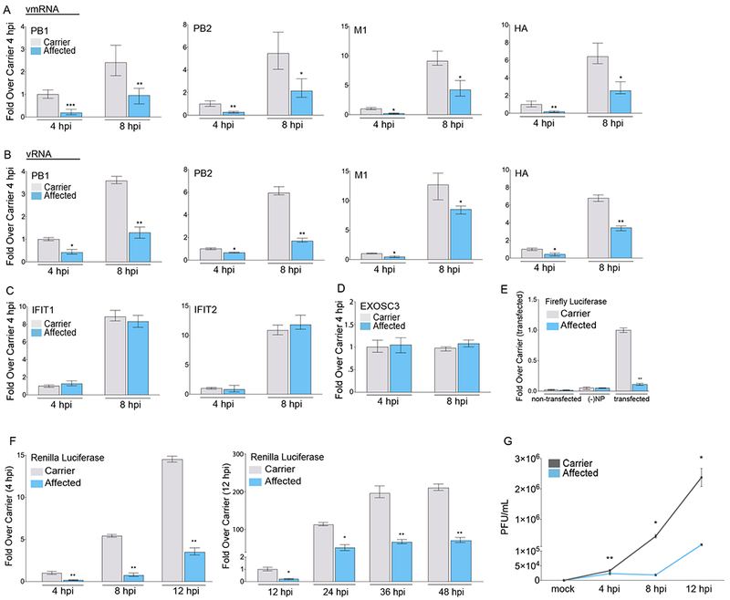
(A-C) qPCR of IAV viral mRNA (A), RNA (B), and host infection-induced gene mRNA levels (C) in primary dermal fibroblasts isolated from patients with pontocerebellar hypoplasia type I (affected; homozygous mutation – Asp132Ala) or family members (carrier; heterozygous mutation). Cells were infected with the PR8 strain at the indicated time points.
(D) mRNA expression levels for EXOSC3.
(E) IAV minigenome replicon assay in patient primary dermal fibroblasts. Viral polymerase transcribed firefly luciferase levels were normalized to co-transfected renilla luciferase levels.
(F) Renilla luciferase expression in patient primary dermal fibroblasts infected with the IAV PR8-luc at the indicated time points post-infection (single cycle growth – left; multi cycle growth – right).
(G) Viral replication growth curve assayed from supernatants harvested from patient primary dermal infected with IAV PR8 at the indicated time points post-infection.
Statistical analyses between datasets were performed with a two-tailed Student’s t-test, adjusted with a Holm-Bonferroni test for sequential comparisons. For all panels, *p<.05, **p<.005, and ***p<.0005. Error bars indicate SD from triplicate experiments.
See also Figure S3