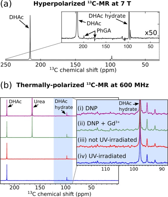
Figure 5.

(a) Representative 13C MR spectrum (sum of 180 spectra) of a hyperpolarized solution containing 40 mM [2-13C]DHAc, 5 mM PhGA, and 6 μM Gd3+ in PBS. The MR acquisition started 16 s after the beginning of the dissolution process with a repetition time of 1 s and a nominal flip angle of 9°. (b) 13C MR spectra of thermally polarized solutions at 300 K and 14.1 T. All samples contained [2-13C]DHAc (40 mM-70 mM) and PhGA (4.5–8.0 mM) in PBS with 10% 2H2O. [13C]urea was added to samples i and ii after dissolution as a reference. These samples were made from frozen beads of 8 M [2-13C]DHAc and 1 M PhGA in water. (i) Frozen beads had been irradiated using a narrowband light source (VisiCure 405 nm) for 200 s and dissolution DNP performed on them. (ii) 1.2 mM Gd3+ had been added to the sample prior to photoirradiation and dissolution DNP. (iii) The frozen beads were dissolved in PBS without photoirradiation or DNP. (iv) The frozen beads were irradiated for 200 s and melted in PBS, but they did not undergo dissolution DNP.
