Figure 1.
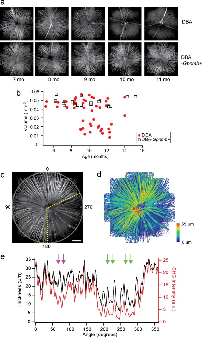
DBA/2J pathologies are detected by SHG imaging. (a) Mosaics of DBA/2J and DBA/2J-Gpnmb+ retinas ex vivo at ages between 7 and 11 months, showing age-dependent loss of the retinal nerve fibers only in DBA/2J mice. (b) The volume of the retinal nerve fibers, displaying significant decrease in DBA/2J mice older than approximately 9 months. (c, d) The integrated SHG intensity and the thickness of the DBA/2J retina (9 months old), respectively. Sectorial degeneration is delineated with yellow dashed lines. Scale bar: 200 μm. (e) The integrated SHG intensity and the thickness versus angle (red and black, respectively), where the regions of decoupling are marked with arrows. The region with low SHG but normal thickness is indicated with magenta arrows.

