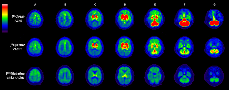
Figure 2.
Multiligand cholinergic PET study in a single PD subject using [11C]-PMP AChE (top row), [18F]-FEOBV VAChT (middle row), and [18F]-Flubatine α4β2 nAChR (bottom row) PET. Columns A-B: all three tracers show uptake in the cortex, with relatively more increased uptake in the anterior frontotemporal cortices. Columns C-D: both [11C]-PMP and [18F]-FEOBV show intense uptake in the striatum and thalamus. [18F]-Flubatine shows intense uptake in the thalamus but only limited uptake in the striatum. Columns E-G: [18F]-FEOBV shows intense uptake in the cerebellar vermis especially while [11C]-PMP has intense uptake in the entire cerebellum. [18F]-Flubatine shows lower and more diffuse uptake in the cerebellum. All three tracers show uptake in the pontine region, with [11C]-PMP > [18F]-FEOBV > [18F]-Flubatine.

