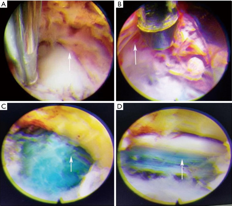
Figure 3.
Intraoperative endoscopic imaging. (A) An existing nerve root is compressed by extraforaminal herniated disc. Arrowhead is pointing to the position of nerve root, same hereinafter; (B) revealing the existing nerve root after removal of the extraforaminal herniated disc; (C) a traversing nerve root is compressed and covered by intracanalicular herniated disc (blue-stained); (D) the traversing nerve root is free after removal of the intracanalicular herniated disc.

