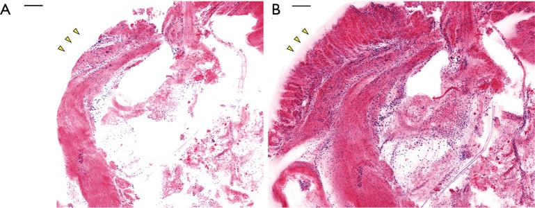
Figure 4.
Result of pseudocolor and imaging without (A) and with (B) the PDMS cover. Mouse stomach tissue was imaged with the confocal microscope system and pseudocolor was applied to resemble H&E histology. With the PDMS holder pressing the tissue down, additional structural information, such as the glandular mucosa, marked with yellow arrows, can be observed. Scale bar: 100 µm.

