
Fig. 2.
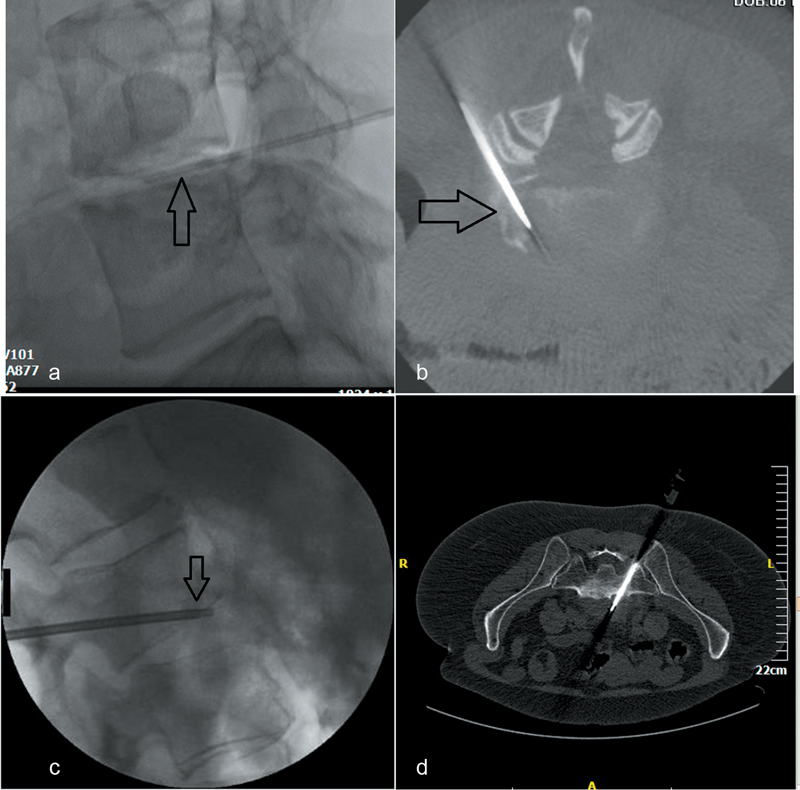
Percutaneous biopsy for spondylodiscitis. ( a ) Lateral fluoroscopic view—needle is inside L4–L5 intervertebral disc (arrow)—biopsy is performed for culture in a patient with suspected spondylodiscitis. ( b ) Cone beam CT axial reconstruction (same patient)—needle is inside L4–L5 intervertebral disc (posterolateral approach). ( c ) Lateral fluoroscopic view (different patient from a, b )—biopsy needle (arrow) is placed inside L2–L3 intervertebral disc trough a transpedicular access crossing the end plate. Sampling is performed both from the end plate and the intervertebral disc. ( d ) Computed tomography axial scan (different patient from a, b, c )—biopsy needle is placed inside the lesion located anterior to the L5–S1 intervertebral disc (transsacral approach).