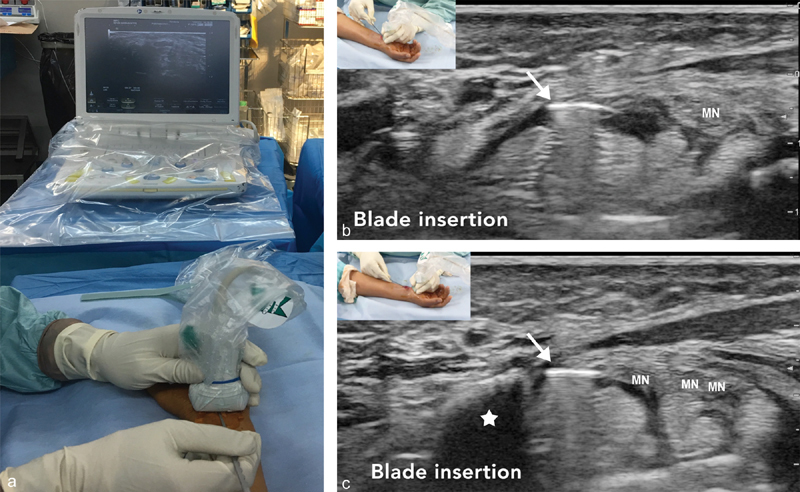
Fig. 3.

( a ) The hook knife is positioned horizontally. Corresponding axial ultrasound images at ( b ) the proximal and ( c ) distal carpal tunnel. Hook knife artifact ( arrow ) under the flexor retinaculum. Hook of Hamate bone (white star). Median nerve (MN) before and after division.
