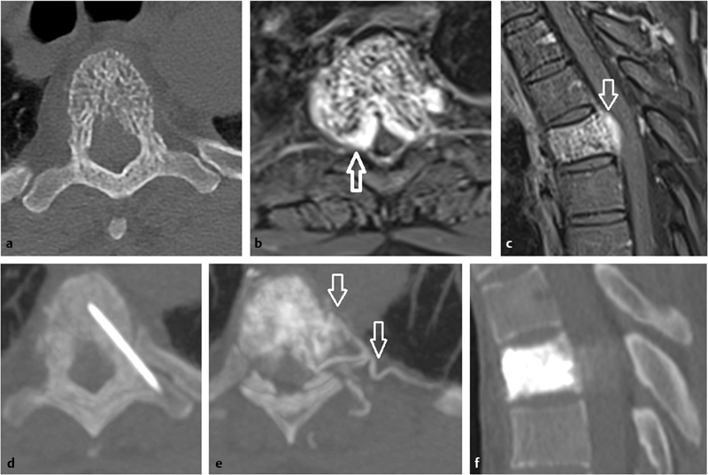
Fig. 2.

A 35-year-old woman with upper back pain due to an aggressive vertebral hemangioma of Th5. ( a ) Axial unenhanced CT scan shows an aggressive vertebral hemangioma of T5 with typical sparse and coarsened vertebral trabeculae. ( b, c ) Axial and sagittal fat-suppressed T1-weighted contrast-enhanced MR images identify a large extraosseous and epidural component ( white arrows ) leading to a severe thoracic canal stenosis. ( d ) Axial unenhanced intraprocedural CT scan shows the position of an 18-gauge needle in the vertebral body. ( e ) Axial intraprocedural CT scan during a venogram identifies the extensive vascularity of the hemangioma inside the vertebra and the surrounding soft tissue ( arrows ). ( f ) Postprocedure sagittal CT scan demonstrates cement filling the vertebral body.
