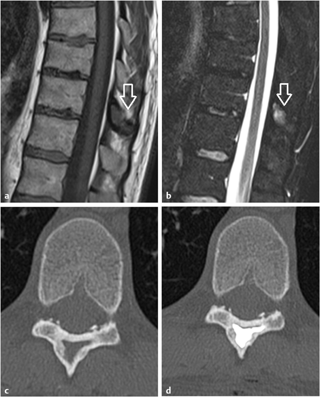
Fig. 3.

A 28-year-old man with persistent back pain caused by fibrous dysplasia of the spinous process of T11. ( a, b ) Unenhanced sagittal T1- and T2-weighted MR images demonstrate fibrous dysplasia of T11 as a hypo-T1 hyper-T2 lesion ( white arrows ) of the spinous process. ( c ) Axial unenhanced CT scan of T11 shows endosteal scalloping, opaque ground-glass abnormal bone, and a peripheral thick reactive bone. ( d ) Postprocedure CT scan demonstrates cement filling the lytic portion of the fibrous dysplasia.
