Figure 1.

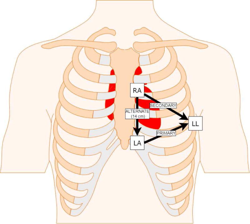
Location of Left Arm (LA), Left Leg (LL), and Right Arm (RA) electrodes placement for the 3-lead ECG to mimic the primary, secondary, and alternate sensing vectors of the S-ICD. Lead RL was placed on the lower right leg.

Location of Left Arm (LA), Left Leg (LL), and Right Arm (RA) electrodes placement for the 3-lead ECG to mimic the primary, secondary, and alternate sensing vectors of the S-ICD. Lead RL was placed on the lower right leg.