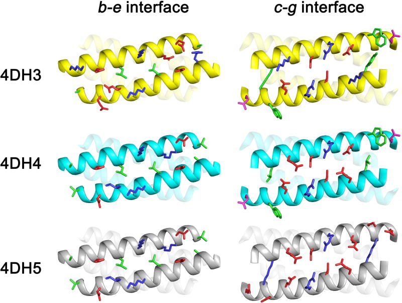
Figure 8.
Structural differences among the designed models of 4DH3, 4DH4 and 4DH5. The main chain is presented as cartoon, residues in the b and e (on the left) and c and g (on the right) positions as sticks. Hydrophobic, polar, negative- and positively-charged residues are coloured in green, magenta, red and blue, respectively. Helices 2 and 3 (on the left) and helices 3 and 4 (on the right) are transparent to allow a clearer view of the interfaces.

