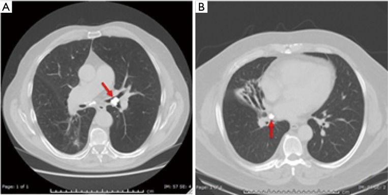
Figure 2.
Common CT imaging findings in patients with broncholithiasis. (A) Axial chest CT image demonstrates a broncholith (arrow) in the left mainstem bronchus without parenchymal involvement; (B) axial chest CT image of a different patient demonstrates a broncholith (arrow) at the origin of the right middle lobe bronchus with associated bronchiectasis and parenchymal atrophy.

