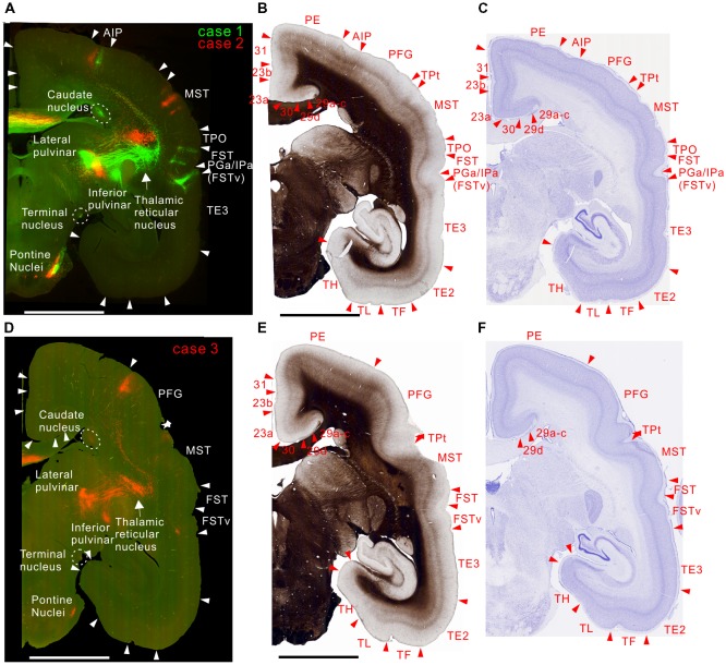
Figure 5.
Brain section images at levels of rostral from the injection sites, utilizing the same format as in Figure 2. (A) Fluorescent section image from case 1 (green) and 2 (red). (B) Image of the corresponding myelin-stained section. (C) Images of the corresponding Nissl substance-stained section. (D–F) Images of brain sections from case 3.

