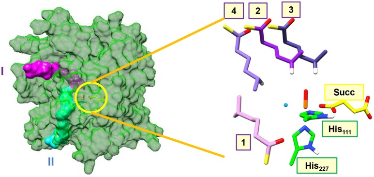
Figure 2.
Substrate entrance channels I (in purple) and II (in cyan) into the active site of the halogenase domain of HctB. The yellow circle highlights the active site of the halogenase in the protein. The right-hand-side of the figure gives an overlay of the four substrate binding positions, where we only show the hexanoyl group and highlight the transferring hydrogen atom in white. Substrate binding position 1 is located in entrance channel II and substrate binding position 2, 3, and 4 are bound in entrance channel I.

