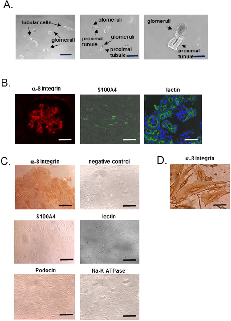
Figure 2.
Characterization of primary culture cells. (A) The observation of sieving sample under phase-contrast microscope. The sieving sample contains glomeruli and other cells such as tubular cells. Right panel shows sieving sample containing glomeruli connected to proximal tubules. Scale bar, 20 µm (left and middle panel), 8 µm (right panel). (B) Immunofluorescent staining of α-8 integrin (mesangial cell marker) and S100A4 (fibroblast marker) in kidneys of C57BL/6 J mice. A glomerulus contains cells with positive staining for α-8 integrin, and some cells with positive staining for S100A are observed in glomeruli and tubulo-interstitial cells. Right panel shows proximal tubular cells and their cells invaginated into parietal layer of Bowman’s capsule with the positive staining for lotus tetragonolobus lectin. Scale bar, 20 µm (left panel), 7 µm (middle and right panel). (C) Immunohistochemistry of α-8 integrin, S100A4 with the negative control, podocin (podocyte marker), Na-K ATPase (tubular cell marker) and fluorescence of lotus tetragonolobus lectin (proximal tubular cell marker) in mesangial primary culture cells. All most of the primary cultured cells exhibit positive staining for α-8 integrin but negative staining for the other markers. Scale bar, 20 µm. (D) High magnification of α-8 integrin staining. The cells after primary culture included an irregular shape and flattened-cylinder-like cell bodies, typical of mesangial cells. Scale bar, 4 µm.

