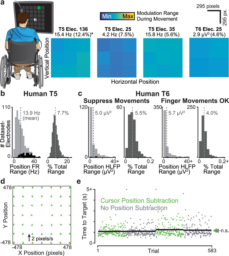
Figure 5.
BMI cursor position correlates in human motor cortex are small. (a) An analysis similar to that of Fig. 2 was performed using motor cortical data from two human clinical trial participants performing a Grid Task with a BMI cursor. Trials were grouped by dividing the target locations within the workspace into nine (participant T5) or four (participant T6) tiles. The plot shows, for four example electrodes, trial-averaged neural activity immediately prior to target selection. Data are from datasets T5.2016.10.13 and T6.2014.07.02. Note that for T6, the principal BMI control signal was high frequency LFP power (HLFP), rather than spike rate. (b) Data from ‘T5’, who has spinal cord injury and is unable to volitionally move his arms, presented as in Fig. 2c. (c) Similar analysis for ‘T6’, who has ALS and was still able to volitionally move her hands. We collected data in two different behavioral contexts: in the movement suppressed context (left), we asked T6 to avoid moving her hand. In the regular context (right), she was free to (and did) make finger movements during BMI use. (d) We performed a closed-loop decoder comparison with and without Cursor Position Subtraction (dataset T5.2017.06.28). This panel shows the effect of Cursor Position Subtraction on cursor velocity as in Fig. 1d. (e) Trial-by-trial closed-loop Radial 8 Target Task performance, shown as in Fig. 1b. Two points with values of >5 s are drawn at the plot ceiling. Arrows show each decoder’s mean time to target.

