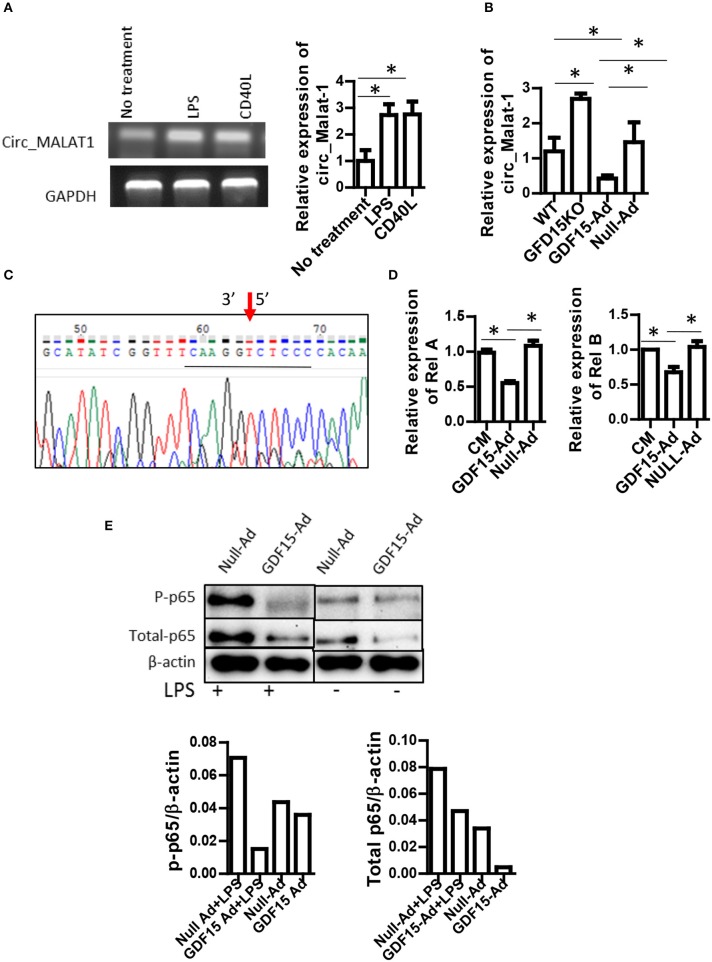
Figure 5.
GDF15 reduced circ_Malat-1 expression and inhibited the NFκB pathway. (A) circ_Malat-1 expression in DCs. BM derived DCs were cultured from WT mice and treated with 50 ng/ml CD40 L or 10 ng/ML LPS for 24 h.Untreated DCs were used as control. RNA was extracted and circ_Malat-1 expression was detected by RT-PCR. Left: regular PCR, representative images for n = 3; Right: qPCR, n = 3, One way ANOVA was conducted for statistical analysis *P < 0.05. (B) GDF15 negatively regulated circ_Malat-1 expression. The expression of circ_Malat 1 was detected in GDF15 KO DCs, WT DCs or GDF15-Ad or control Null-Ad treated DCs by qRT-PCR. n = 3, One way ANOVA was conducted for statistical analysis *P < 0.05. (C) Representative DNA sequence from DNA sequencing of RT-PCR products. circ_Malat-1 was amplified using divergent primers and the PCR products was then subjected to DNA sequencing using forward primer of circ_Malat-1. Arrow: pointing to the junction of circ_Malat-1. The sequencing containing the circ_Malat-1 conjunction sequence gcatatcggttt caaggt ctcc ccacaa was presented. The central conjunction sequence was underlined. (D) The expression of Rel A and Rel B by qRT-PCR. DCs were treated with GDF5-Ad, or Null-Ad. The expression of Rel A and Rel B were measured by qRT-PCR 48 h after infection. One way ANOVA was conducted for statistical analysis n = 3 *P < 0.05. (E). GDF15 inhibited the NFkB signaling pathway. Phosphorylated p65 and total p-65 protein was detected by western blotting using phosphorylated p65A Abs and p65 Abs. Representative of image of western blotting (upper) and relative quantity of protein (low) from n = 4 experiments. Samples treated with LPS and samples without LPS treatment were loaded separately by other samples for PAGE.

