Figure 1.
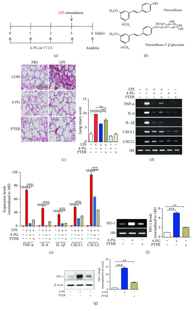
4-PG prevents LPS-induced acute lung injury and upregulates HO-1 expression. (a) The scheme depicts the experimental protocol used to assess the protective effect of pterostilbene 4′-glucoside (4-PG) and pterostilbene (PTER) on LPS-induced ALI. 10-week-old mice were injected with 4-PG (10 mg/kg, i.p.) and PTER (10 mg/kg, i.p.) for 4 days prior to intranasal administration of LPS (2.5 mg/kg) for 24 h. (b) Chemical structures of 4-PG and PTER. (c) Lung sections were stained with hematoxylin and eosin (H&E) for morphological evaluation, and the representative lung sections of each group are shown. Scale bar = 100 μm. (left). Quantitative analysis of histologic lung section by lung injury score for six experimental groups. The score generates the average of two independent investigators (right). (d, e) The mRNA expression of proinflammatory cytokines and chemokines (TNF-α, IL-6, IL-1β, CXCL1, and CXCL2) in lung tissues was detected by RT-PCR. Furthermore, mRNA and protein levels of HO-1 were assessed by RT-PCR (f, left: HO-1mRNA levels, right: quantification of the relative band density) and Western blotting (g, left: HO-1 protein levels, right: quantification of the relative band density) from lung tissues, respectively. 18S and β-actin were used as internal controls. Data were expressed as mean ± SD (n = 5 per group); ∗∗p < 0.01 and ∗∗∗p < 0.001. Comparisons were made by one-way ANOVA with Turkey post hoc tests.

