Figure 3.
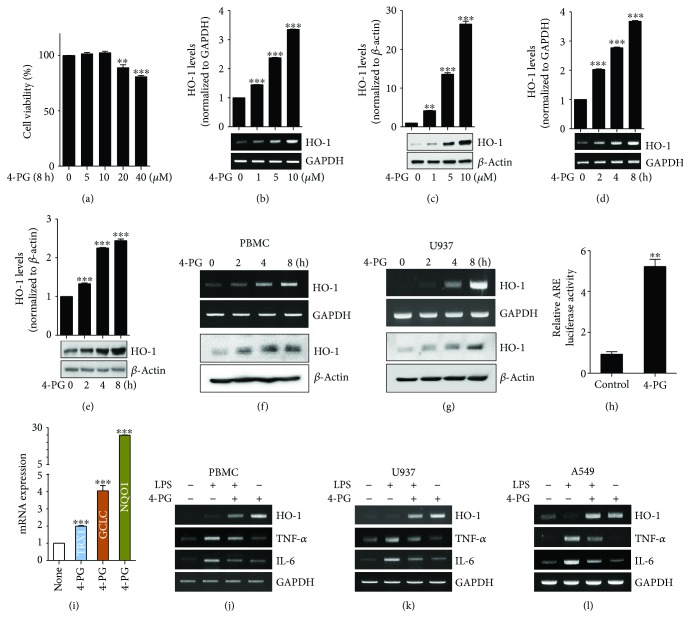
4-PG induces HO-1 mRNA and protein expression in macrophages and lung epithelial cells. (a) RAW 264.7 cells were treated with 4-PG at various concentrations (0, 5, 10, 20, and 40 μM) for 8 h, and cell viability was determined by MTT assay. To evaluate the beneficial effect of 4-PG on HO-1 induction, cells were treated with 4-PG (0, 1, 5, and 10 μM) at the indicated concentrations for 8 h. The mRNA and protein levels of HO-1 were measured by RT-PCR (b) and Western blotting (c). RAW 264.7 cells were treated with 4-PG (10 μM) at the indicated time points (0, 2, 4, and 8 h). The mRNA and protein levels of HO-1 were determined by RT-PCR (d) and Western blotting (e). (f and g) PBMC and U937 cells were treated with 4-PG at the indicated concentrations (0, 1, 5, and 10 μM) for 8 h. The mRNA and protein levels of HO-1 were determined by RT-PCR (top) and Western blotting (bottom). GAPDH and β-actin were used as internal controls. (h) RAW 264.7 cells were cotransduced with a pCignal Lenti-ARE reporter and pCignal Lenti-TK-Renilla. After treatment with 4-PG, luciferase activity was analyzed. The expression levels obtained from pCignal Lenti-ARE reporter-transduced cells without 4-PG treatment were normalized to 1. (i) RAW 264.7 cells were treated with 4-PG (10 μM) for 8 h. Several antioxidative genes including TRX1, GCLC, and NQO1 were measured by RT-qPCR. (j and k) PBMC and U937 cells were pretreated with 4-PG (10 μM) for 6 h followed by the stimulation of LPS (100 ng/ml) for another 4 h. (l) A549 cells pretreated with 4-PG (10 μM) for 4 h and then stimulated with LPS (10 μg/ml) for 6 h. The mRNA levels of HO-1, TNF-α, and IL-6 were determined by RT-PCR. Data were expressed as mean ± SD (n = 5 determined in five independent experiments). One-way ANOVA with Turkey post hoc tests were performed; ∗p < 0.05, ∗∗p < 0.01, and ∗∗∗p < 0.001 vs. the vehicle control group.

