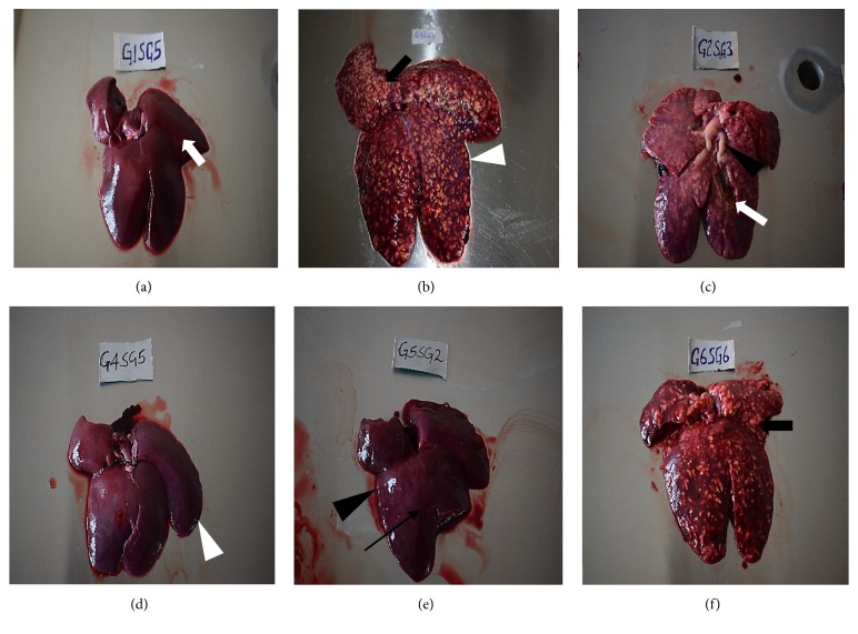
Figure 2.
Hepatic lesions at termination of the drug efficacy study in experimentally induced coccidiosis infection. (a) Normal liver with normal architecture of incised section (white arrow) from negative control group, (b) liver with hepatomegaly manifested by diminished sharp edges (white arrow head) with raised multinodular lesions due to hepatic coccidiosis from positive control group, (c) markedly enlarged liver with multinodular whitish-yellow lesions and distended bile duct (arrow head), incised section with greenish-yellow material (white arrow) from amprolium treatment group, (d) liver from diclazuril treatment group without any significant lesion, note the sharp edges (white arrow head), (e) slightly enlarged liver (loss of sharp edges, black arrow head) with tiny whitish-yellow fibrotic spots after healing (black arrow) from sulphachloropyrazine group, and (f) enlarged liver with raised multinodular whitish-yellow lesions from trimethoprim-sulphamethoxazole group.

