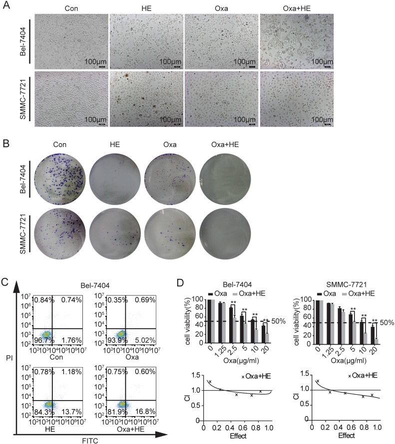
Fig 3.
Effects of Oxa, HE, and Oxa combined with HE in HCC cell lines. (A-D) Bel-7404 and SMMC-7721 cells were incubated with Oxa (10 μg/ml), HE (12mg/ml), or Oxa (10 μg/ml) combined with HE (12mg/ml) for 24h. The IC50 of HE was measured by CCK8 assay (Figure S1). (A) The morphological changes were observed by inverted microscope. (B) Clone formation capacity of Bel-7404 and SMMC-7721 cells was assessed by the clone formation assay. Cells were stained with 0.1% crystal violet after fixed. (C) Representative early apoptotic cells were detected by FCM following Annexin V-FITC/PI staining in Bel-7404 cells. The lower right quadrant of the cross represented early apoptotic cells (%). (D) CCK-8 was performed to assess the cytotoxic effect of Oxa, HE, and Oxa combined with HE. The combination index (CI) value represented the mutual effect of Oxa and HE. CI lower than 1.0 represented synergistic effect. Each bar represented means± SD from three independent experiments. *, P<0.05, **, P<0.01 as analyzed by Student t test.

