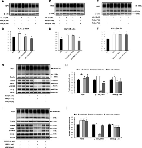
Figure 2.
LCA increased AQP2 protein expression via cAMP-PKA signaling pathway. Primary IMCD suspensions were pretreated with DMSO (vehicle), H89 (20, 30 μM), MDL-12330A (10, 100 μM), or tolvaptan (10−11, 10−10 M) for 30 minutes and then treated with LCA (10 μM) or CDCA (100 μM) for 6 hours. Cell lysates were subjected to Western blot analysis for AQP2 (A–F), p-CREB (ser133), CREB, p-GSK3β (ser9), GSK3β or β-actin (G–J); n=6 biologically independent samples in each groups. Data are shown as mean±SEM; *P<0.05 compared with CTL; #P<0.05 compared with either LCA or CDCA. CTL, control; MDL, MDL12330A; Tol, tolvaptan.

