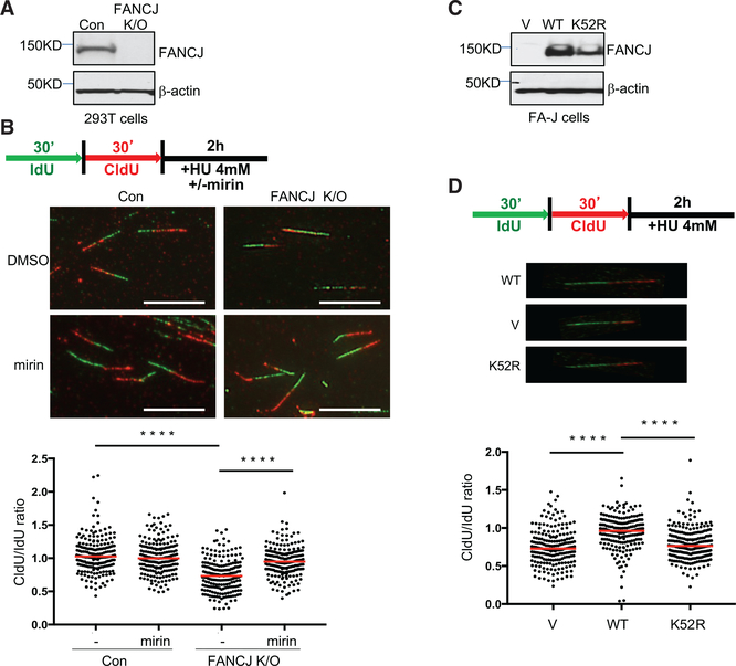
Figure 1. FANCJ and Its Helicase Activity Protect Nascent DNA at Replication Forks from MRE11-Dependent Degradation.
(A) Western blot analysis with the indicated antibodies (Abs) of lysates from control and FANCJKO 293T cells.
(B) Schematic, representative images, and quantification of the CldU/IdU ratio after HU treatment with or without mirin.
(C) Western blot analysis with indicated Abs of lysates from FANCJ-null FA-J cells complemented with vector (V), wild-type (WT), or a catalytically inactive FANCJ (K52R) mutant.
(D) Schematic, representative images, and quantification of the CldU/IdU ratio after HU treatment. Each dot represents one fiber.
For each analysis, at least 200 fibers are quantified from two independent experiments. Red bars represent the median. Statistical analysis according to two-tailed Mann-Whitney test; ****p < 0.0001. Scale bars, 10 μm.

