Fig. 3.
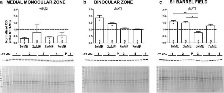
Time-dependent effect of ME on cortical area-specific vMAT2 expression levels. Western blot OD values are illustrated as the ratio of the expression levels of each ME group relative to the expression of the AMC group to judge the effect of post-ME recovery time. Below each graph, representative Western blot bands are depicted, as well as the expected molecular weight (~ 75 kDa) and an example total protein stain. The different conditions are indicated with numbers above the respective bands: 1 = 3wME, 2 = 5wME, 3 = AMC, 4 = 7wME and P = region-specific tissue sample pool. a At 1 week post-ME (1wME), the expression of vesicular monoamine transporter 2 (vMAT2) in the medial monocular zone (Mmz) was significantly reduced compared to the AMC mice (dashed line) and the vMAT2 expression levels remain low, even after 7 weeks. b In the binocular zone (Bz), a significant ME-effect was detected in 1wME and 3wME mice, showing increased vMAT2 expression levels immediately after ME. c In the primary somatosensory barrel field (S1BF), vMAT2 expression was increased in 1wME mice compared to AMCs, and was significantly higher in 1wME and 3wME mice compared to 5wME mice. The number of animals (n) is represented in the bars. Significant pair-wise differences between ME mice and the AMC group are indicated with a ‘*’ within the corresponding bar. Differences between ME conditions due to post-ME recovery time are indicated as a ‘*’ above the respective ME conditions. *P < 0.05, **P < 0.01

