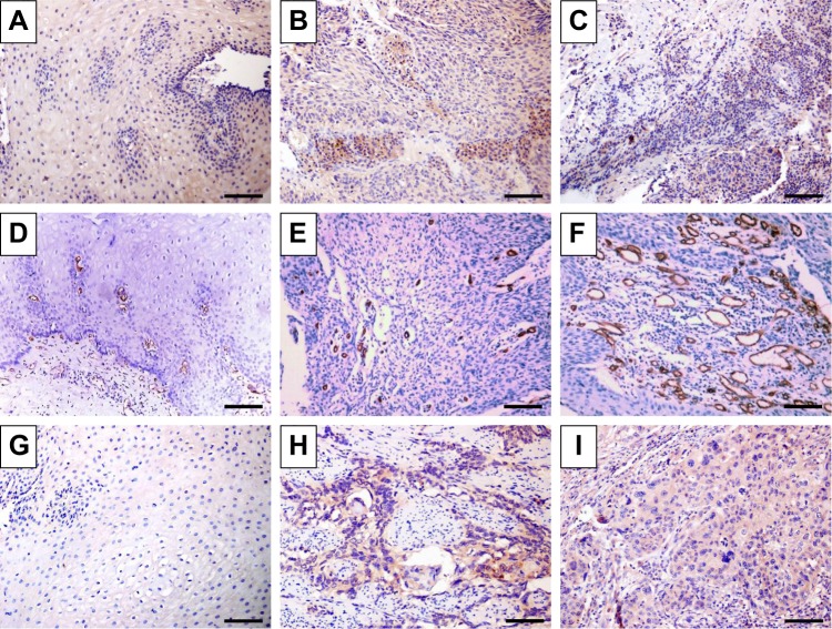
Figure 1.
Immunohistochemical staining of normal and esophageal cancer specimens in which antibodies to AIP1 (A–C), CD34 (D–F), and VEGFR2 (G–I) were used.
Notes: Representative immunostaining images of (A, D, G) normal adjacent tissues and (B, C, E, F, H, I) ESCC tumor tissues. (B and C) Distribution of AIP1 in ESCC tumor tissues revealed diffuse staining of membranes and cytoplasm of ESCC tumor tissues. (C) Low density of AIP1 located in ESCC tissues. (D–F) Immunohistochemical staining of CD34, which was used to mark endothelial cells and to evaluate MVD in different tissues. (E) Low MVD in ESCC tissues. (F) High MVD in ESCC tissues. (H and I) Different distribution of VEGFR2 in ESCC tumor tissues. Scale bar=100 µm.
Abbreviations: AIP1, ASK1-interacting protein-1; ESCC, esophageal squamous cell carcinoma; MVD, microvessel density; VEGFR2, vascular endothelial growth factor receptor 2.

