Figure 3.

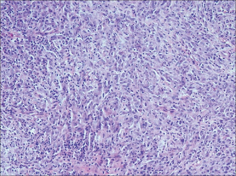
Photomicrograph of the fibroinflammatory polyp shows proliferating myofibroblasts, and inflammatory infiltrate of lymphomononuclear cells (H and E, ×20)

Photomicrograph of the fibroinflammatory polyp shows proliferating myofibroblasts, and inflammatory infiltrate of lymphomononuclear cells (H and E, ×20)