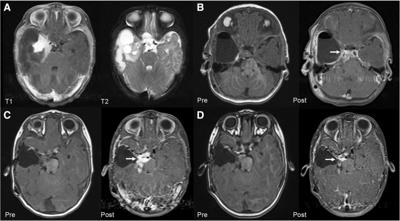
Fig. 1.
Radiologic findings. a (T1-Weighted Postcontrast and T2-Weighted Axial Magnetic Resonance Imaging), Large right inferomedial temporal solid-multicystic mass with a postcontrast enhancing component. b (T1-Weighted Pre and Postcontrast Axial Magnetic Resonance Imaging), Three-month postoperative leptomeningeal spread involving the upper brainstem. c (T1-Weighted Pre and Postcontrast Axial Magnetic Resonance Imaging), Eight-month postoperative progression of leptomeningeal involvement despite standard chemotherapy. d (T1-Weighted Pre and Postcontrast Axial Magnetic Resonance Imaging), Fourteen-month postoperative decrease in residual tumor after 6 months of BRAF-MEK inhibitor therapy

