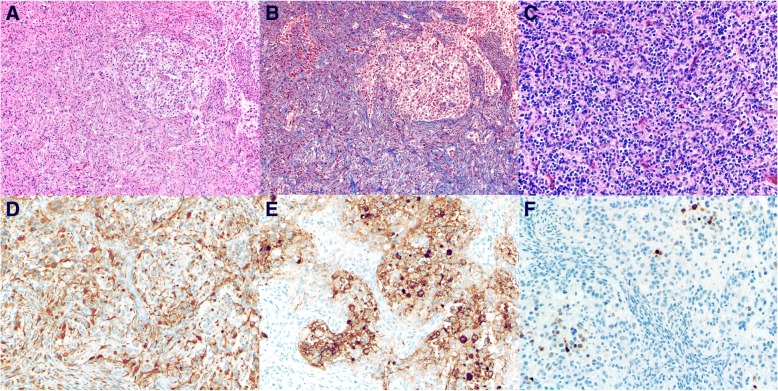
Fig. 2.
Histologic findings. a, Astrocytic and neoplastic neuronal tumor cell components (hematoxylin-eosin, × 100). b Prominent desmoplastic stroma (Masson’s trichrome, 100x). c Focal poorly differentiated neuroepithelial (small cell) component (hematoxylin-eosin, × 200). d Glial fibrillary acidic protein immunostain highlighting the astrocytic tumor cell component (× 200). e Synaptophysin and f Neu-N immunostain highlighting the neoplastic neuronal tumor cell component (× 200)

