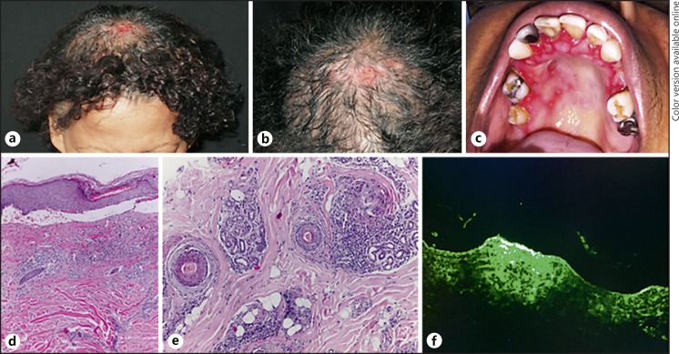
Fig. 1.
a, b Area of cicatricial alopecia in the parietal region with exulcerations. c Erosions on the hard palate and desquamative gingivitis. Histopathology of the scalp: subepidermal vesicle with influx of lymphocytes and eosinophils in the papillary dermis (d); concentric fibroplasia in the terminal follicles with lichenoid infiltrate with lymphocytes, plasma cells, and rare eosinophils (e). f Direct immunofluorescence with salt-split test of the scalp positive for IgG antibody on the epidermal side of the blister.

