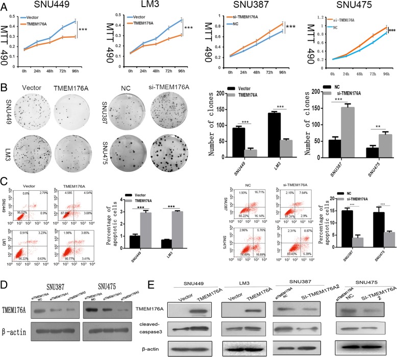
Fig. 3.
Effect of TMEM176A on HCC cell proliferation and apoptosis. a Growth curves represent cell viability analyzed by the MTT assay in TMEM176A re-expressed and unexpressed LM3 and SNU449 cells, as well as in SNU387 and SNU475 cells before and after knockdown of TMEM176A. Each experiment was repeated in triplicate. *P < 0.05, ***P < 0.001. b Colony formation results show that colony numbers were reduced by re-expression of TMEM176A in LM3 and SNU449 cells, while they were increased by knockdown of TMEM176A in SNU387 and SNU475 cells. Each experiment was repeated in triplicate. Average number of tumor clones is represented by bar diagram. *P < 0.05, ***P < 0.001. c Flow cytometry results show induction of apoptosis by re-expression of TMEM176A in LM3 and SNU449 cells, while reduction of apoptosis was found after knockdown of TMEM176A in SNU387 and SNU475 cells. *P < 0.05,***P < 0.001. d Knockdown of TMEM176A in SNU387 and SNU475 cells by siRNA. TMEM176A expression was examined by Western blots. SiTMEM176ANC: SiRNA for TMEM176A negative control; SiTMEM176A1: SiRNA for TMEM176A set1; SiTMEM176A2: SiRNA for TMEM176A set2. e Western blots show the effects of TMEM176A on the levels of cleaved caspase-3 expression in LM3, SNU449, SNU387, and SNU475 cells. VECTOR: control vector, TMEM176A: TMEM176A expressing vector, β-actin: internal control, NC: siRNA negative control

