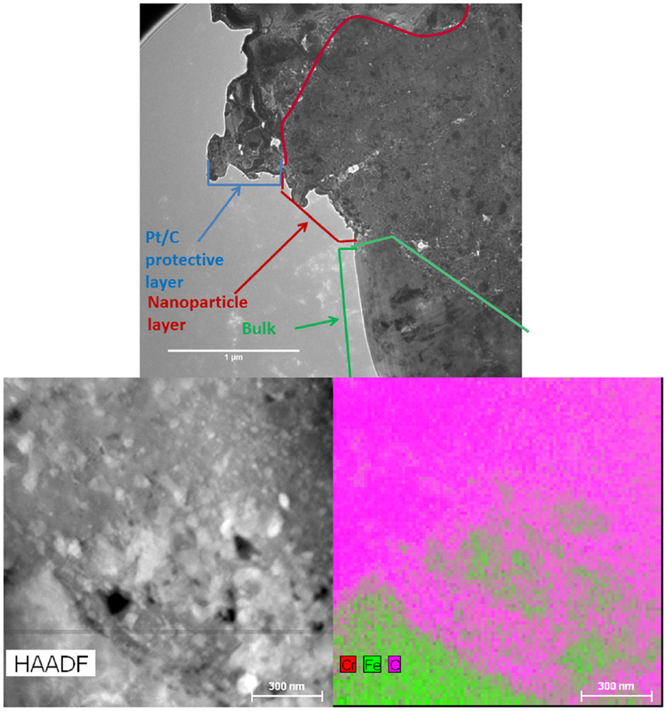
Fig. 6.
Top: cross section view of the nanoparticle layer and bulk material interface (1 μm scale bar). Bottom left: high angle annular dark field TEM image of the interface between the nanoparticle layer and the bulk material. Bottom right: combined energy dispersion X-ray spectroscopy map of the interface showing that the nanoparticles are primarily composed of iron, chromium, and oxygen (300 nm scale bar).

