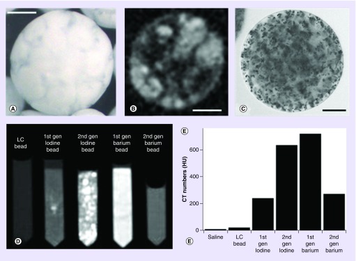
Figure 2. . Appearance and properties of early radiopaque bead prototypes.
(A) Optical image of Lipiodol-loaded bead. Note the marbled appearance caused by the separation of the oil and water phases within the microsphere structure. (B) Micro-CT image of the Lipiodol-loaded bead highlighting the oil (light) and water (dark) phases in the internal structure. (C) Optical image of a barium sulfate-loaded bead clearly showing the precipitated barium sulfate particulates within the hydrogel structure. Scale bars are 100 μm for (A–C). (D) Beads suspended in tubes imaged with CT shown with window settings chosen to display maximum contrast. (E) Corresponding quantitative CT numbers calculated for saline, unloaded LC beads, first generation and second generation of iodine- and barium-loaded beads.
CT: Computed tomography.

