
Figure 4.
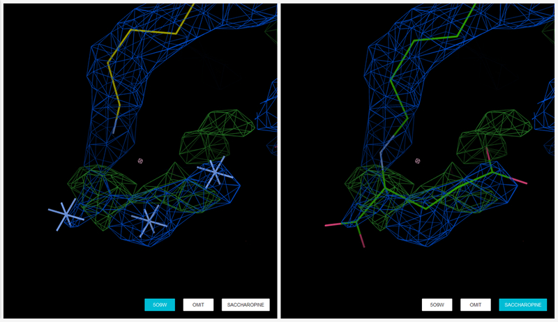
A more speculative interpretation can be provided by submitting the files to Molstack (http://molstack.bioreproducibility.org). Due to disorder and weak density, the possible modification of the lysine was marked as unknown atoms in the deposited structure (left), but using Molstack (http://molstack.bioreproducibility.org/project/view/OJM60NCMQF1VMKW0IBUF/), Kluza et. al [99] provided a probable interpretation that the modified lysine is a saccharopine resulting from the crystallization conditions (right). 2mFo – DFc maps are displayed in blue contoured at a level of 1.0 rmsd. mFo - DFc difference maps are contoured at the 3.0 rmsd level in green (positive) and red (negative).