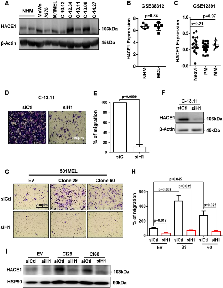
Fig. 1.
HACE1 expression is maintained in melanoma cells and promotes melanoma cell migration. a Western blot analysis of HACE1 and actin protein levels in melanoma cell lines, (MeWo, A375, 501MEL) short-term melanoma cell cultures (C-10.12, C-12.34, C-13.11, C-13.08, C-14.27) and primary human melanocytes (NHM). b Analysis of GSE38312 data sets for HACE1 expression in pairs of cultured cutaneous melanocytes (NHM) and melanoma cells (MC). c Analysis of GSE12391 data sets for HACE1 expression in metastatic (MM) (n = 5) or primary melanomas (PM) (n = 23) compared with that in nevi (n = 18). In b and c, the mean ± SD is represented, and the statistical analysis was performed using t-test with Welch’s correction. d Boyden chamber migration assay of a short-term melanoma cell culture (C-13.11) treated with control (siCtl) or HACE1 (siH1) siRNA. Representative images are shown. e Quantification of three independent experiments. The results are expressed as the percentage of cells in control conditions (mean ± SD; n = 3). f Western blot analysis of C-13.11 cells following siRNA treatment. g Migration assay of 501MEL clones with forced expression of HACE1 (clones 29 and 60). h Quantification of three independent experiments. i Western blot analysis showing HACE1 expression

