Fig. 1.
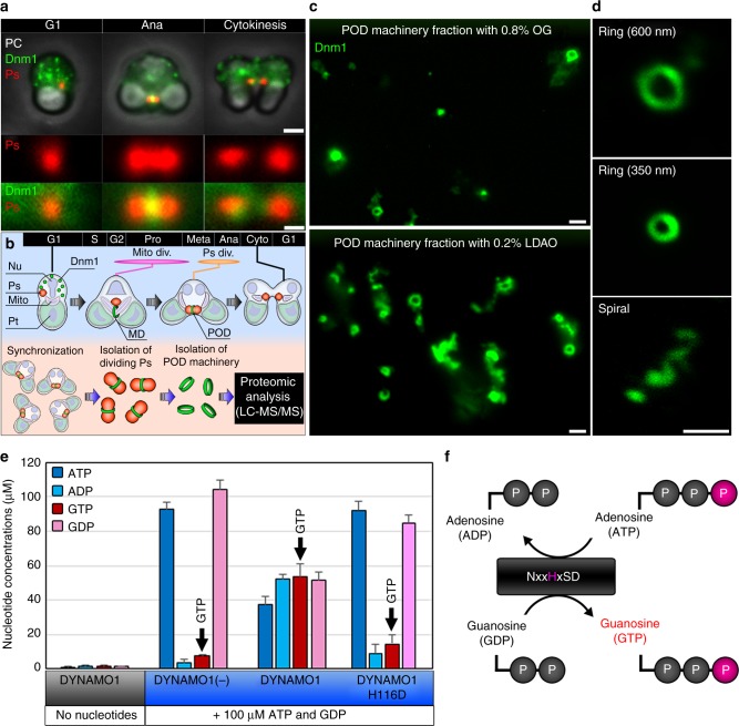
Proteomic analysis of POD machinery and identification of DYNAMO1. a Phase contrast (PC) and immunofluorescence images of C. merolae cells during G1 phase, anaphase, and cytokinesis. Ps peroxisome (anti-catalase antibody), Dnm1 (anti-Dnm1 antibody). b Schematic representing isolation and proteomic analysis of POD machinery. Nu cell nucleus, Mito mitochondrion, Pt plastid, Mito div. mitochondrial division period, Ps div peroxisomal division period. c Upper panel shows the 0.8% OG-treated POD machinery fraction and the lower panel shows the 0.2% LDAO-treated POD machinery fraction. d Typical structures of isolated POD machinery stained with the anti-Dnm1 antibody. e LC–ESI–MS/MS analysis of the nucleoside diphosphate kinase activity of recombinant DYNAMO1. f Schematic representing a working model of nucleoside diphosphate kinase. Data in e are means ± s.d. (n = 3). Scale bars: 1 μm (a, upper panels); 500 nm (a, lower panels; c, d)

