Fig. 5.
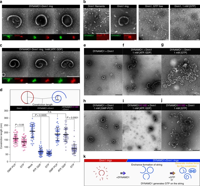
Structure of DYNAMO1-Dnm1 string. a Typical structure of DYNAMO1-Dnm1 strings under GTP-free conditions. Immunofluorescence images of DYNAMO1 (green) and Dnm1 (red) are shown in the respective lower panels. b Typical structure of DYNAMO1-free Dnm1 filaments and strings under GTP-free condition (left, three panels) or in the presence of GTP (right panel). Immunofluorescence images are shown in the lower panels. c Typical structure of DYNAMO1-Dnm1 strings with addition of ATP and GDP. Immunofluorescence images are shown in the lower panels. d Schematic image representing measurement of constriction lengths of the strings. Constriction lengths of the DYNAMO1-free Dnm1, DYNAMO1-Dnm1, and DYNAMO1 H116D-Dnm1 strings under the various conditions (n = 50). e–j Structural dynamics of DYNAMO1-Dnm1 or DYNAMO1 H116D-Dnm1 strings upon addition of ATP and GDP, GTP, or GMP-PCP. k Schematic image representing the dynamics of DYNAMO1-Dnm1 strings. Scale bars: 200 nm (a–c); 500 nm (e–j). Data are means ± s.d. n.s. not significant. P; p-value (unpaired t-test for Dnm1 and DYNAMO1 H116D + Dnm1, Kruskal–Wallis nonparametric ANOVA for DYNAMO1 + Dnm1)

