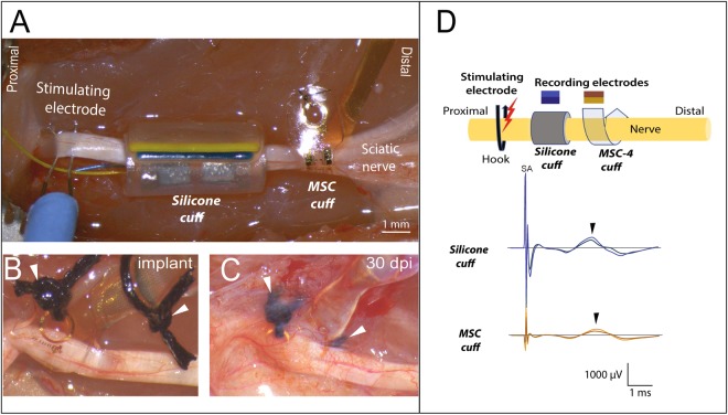
Figure 7.
Sub-chronic recordings form the ScN. (A) In vivo setup showing the placement of the proximal stimulating hook electrode, and side-by-side implantation of the silicone and MSC cuffs electrodes. (B) Photograph of the SMP device immediately after placing and (C) 30 days after implantation, in the latter, the electrode and sutures (arrowheads) are visible through the fibrotic scar. (D) Schematic of the setup with color-coding of the CNAPs recorded from the silicone and MSC devices, both of which recorded the evoked Aδ fiber activity.

