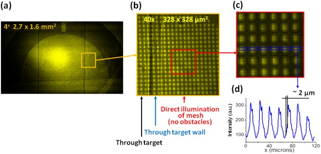
Figure 4.
The image of 1500 lpi mesh obtained on LiF for the evaluation of an instrumental spatial resolution: Panel (a) contains a large field of view photoluminescent image, observed with a 4X microscope objective. Panel (b) shows part of the full image with different elements of target, observed with objective 40X, while panel (c) shows an enlarged crop of this image, in which the diffraction pattern in open areas of the mesh is clearly seen. Panel (d) contains an intensity profile taken from the cropped image.

